Our TMAs
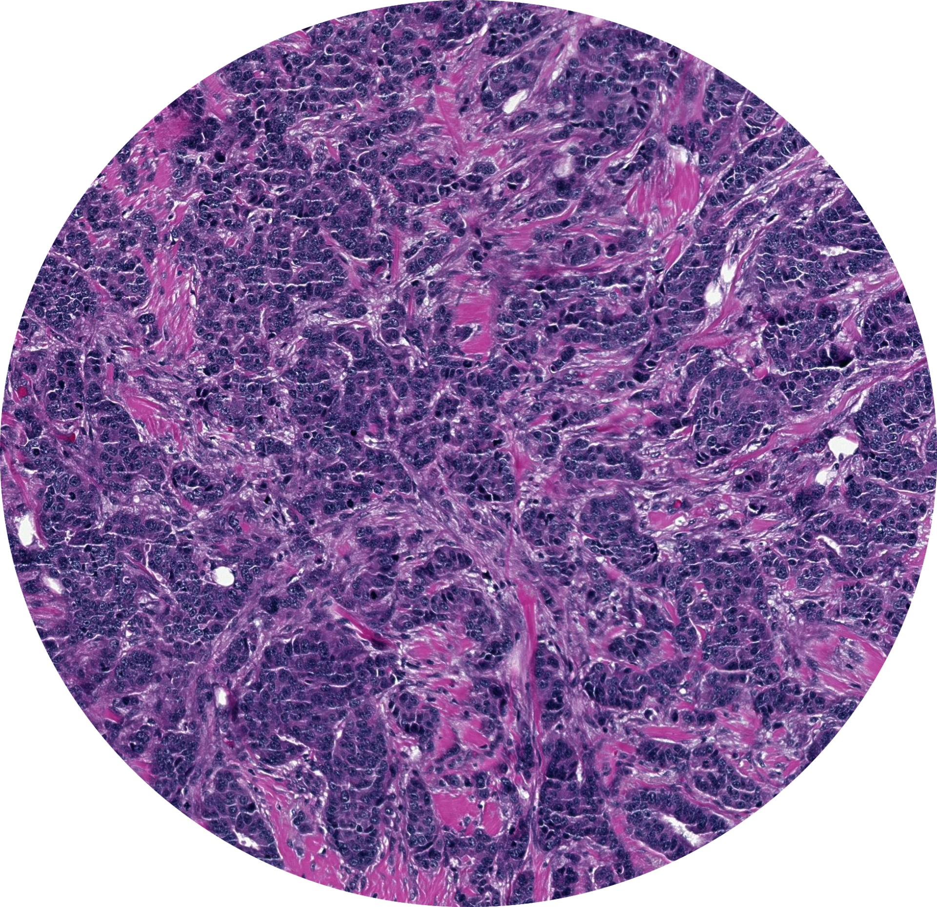
- Suitable for advanced histologic analyses such as immunohistochemistry (IHC) and in situ hybridization (ISH).
- Applications include:
- Assay optimization
- Assay validation
- Daily quality control
- Inter-laboratory comparisons
- Proficiency testing
- Produced from formalin-fixed and paraffin-embedded (FFPE) tissue supplied by clients or tissue carefully selected from trusted commercial suppliers.
- Guaranteed optimal quality and reproducibility thanks to our strict quality control systems.
- Reduced cost by maximizing block yield and minimizing tissue loss thanks to our patented TMA technology.
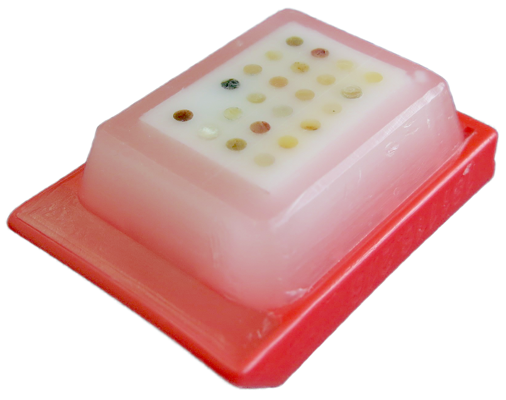
- Built with standard core diameters of 1.0mm, 1.5mm, 2.0mm.
- Custom core sizes are available upon request.
FOR RESEARCH USE ONLY
Customizable
You can specify the number of cores, core diameter and depth, and block height.
We assemble arrays with tissue provided by you or carefully selected from tissue supplied by trusted commercial sources.
Certified
Our tissue is carefully selected from trusted commercial suppliers and validated by a board-certified pathologist. Our strict quality control system ensures optimal quality and reproducibility.
High Yield
With good histologic technique, they yield an estimated 500 or more mounted sections per 5mm cut at 4-5 microns, offering the best price per section.
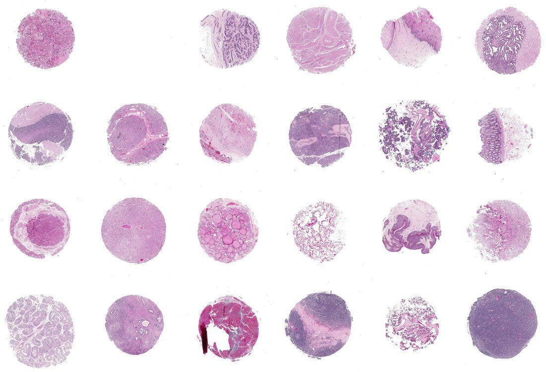
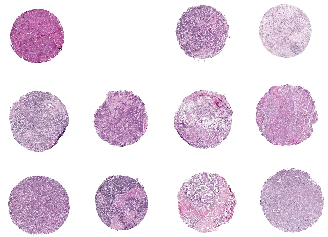
Our Products
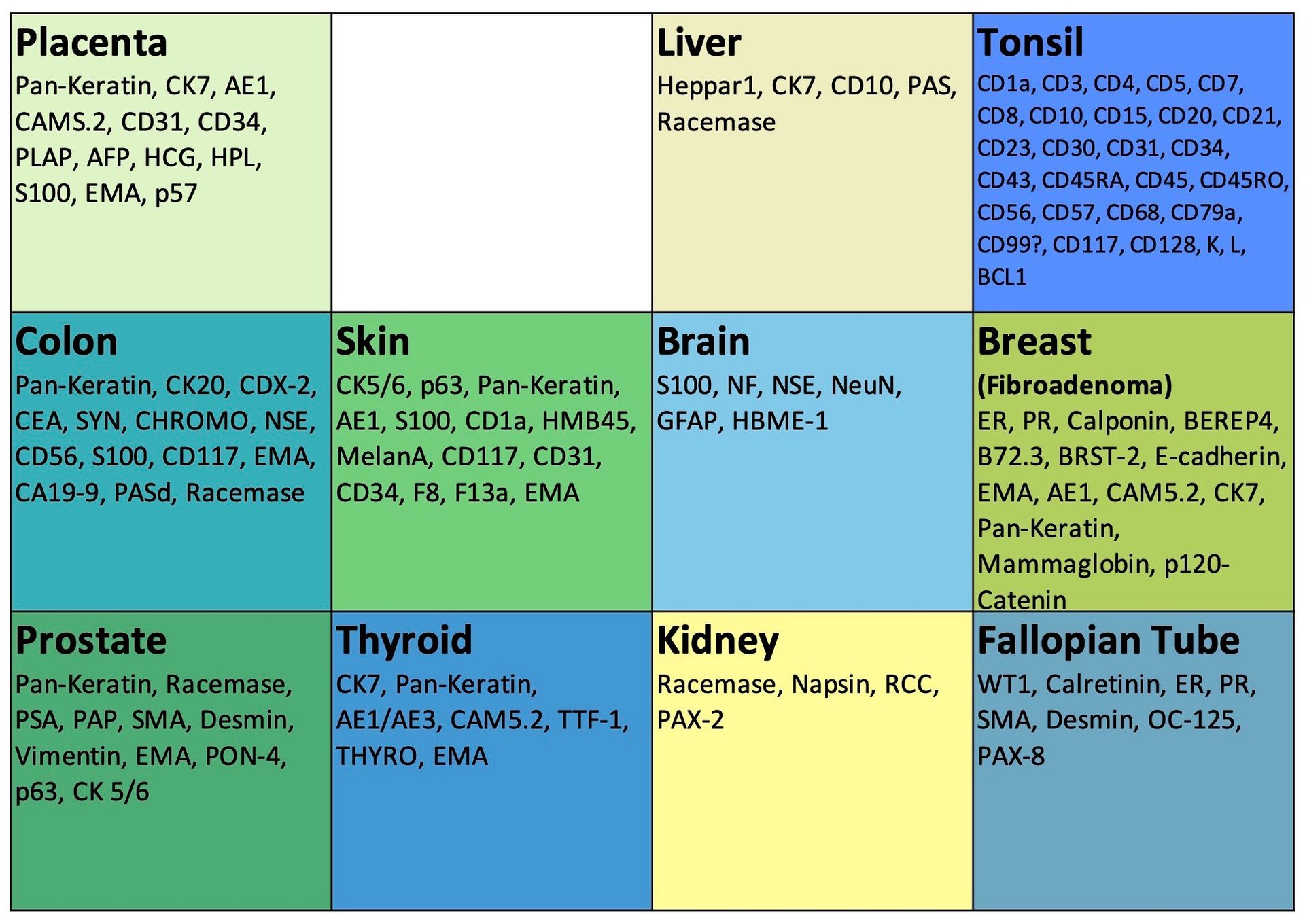
- 11-core or 23-core arrays
- Normal tissue or multiple tumor types
- Example tissue: breast, skin, prostate, colon, brain, thyroid, placenta, pancreas, ovary, salivary gland, testis, thymoma, skeletal muscle, liver. . .